Our People and our Location
To reach our facility you may use the GPS location and enter the address as shown in google maps to reach the correct parking for the facility. For all other purposes please use our mailing address below.
Team Profile
-
Laxmikant (Lax) Saraf, PhD - GENERAL INQUIRY
Director
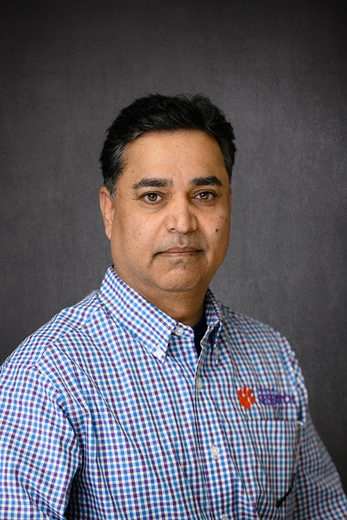
Electron Microscopy Laboratory, Clemson University
lsaraf@clemson.eduEducation:
B.Sc. Physics, Mathematics, Electronics (1990)
M.Sc. Physics, (1992)
Ph.D. Applied Physics (Materials), (1999)Working under the authority of and reporting directly to Clemson University Vice President for Research, Dr. Lax Saraf is currently managing and directing the Electron Microscopy Facility located at Clemson University. He is responsible for supervision EM Facility staff team. Dr. Saraf is a materials scientist/applied physicist in solid state (condense matter) physics with over 25 years of extensive research and development experience in the universities, national lab and industry related to synthesis of advanced materials, oxides/metals/semiconductors thin films. His research on specialized ceramic (oxides) materials was carried out for ferroelectric, microwave, optical, ion transport and superconducting applications. Over the past 18 years, Lax has managed multi-user Electron Microscopy, Thin Film Deposition and Micro-fabrication facilities at Clemson University and US Department of Energy’s national scientific user facilities at Environmental Molecular Sciences Laboratory located at Pacific Northwest National Laboratory (PNNL). Lax was appointed on user facility manager’s council to advice new lab safety implementation, user interactions and communicate strategy changes to managers.
Research Expertise: Electron Microscopy, Fundamentals of Solid Oxide Fuel Cells, Analysis of Li-ion battery cathodes using electron microscopy, Semiconductor material development for Photovoltaics and Solar Cells, Application of Micro/nano-fabrication technology, Nanoscale interface effects in oxides & transport properties, Utilization of microscopic analysis techniques for advanced energy materials, High Frequency Microwave/Magnetic Materials, optical ceramics.
History: Research Assistant, Univ. of Pune, 1992-1997, Research Assistant, University of Maryland, College Park, 1997-1998, visiting researcher from UMD - Corning Applied Technologies, Woburn, MA, 1997-1998. Post-doctoral Scientist at Department of Physics and Department of Materials Engineering at the University of Maryland, College Park, MD, 1999-2001. Research Scientist at PNNL, 2002-2003, Senior Research Scientist at PNNL, 2003-2012. Lab Manager, Micro-fabrication Facility, PNNL, 2002-2008, Lab Manager, Thin Film Deposition Facility, PNNL, 2003-2007, Lab Manager, Electron Microscopy Facility, PNNL, 2008-20012, Director of Electron Microscopy Facility at Clemson University, 2013-Current.
Awards: URSAA Award (2018 - Inaugural class): University Research, Scholarship, and Artistic Achievement Award recognizes Clemson University faculty who have achieved the highest levels of national and international recognition. Faculty selected for URSAAA recognition are lifetime appointees invited to participate in an annual celebration. R&D 100 Award, (top 100 innovations in the world) for the development of Graphene Nanostructures for Li-ion Batteries, 2012, Recognition: Graduate Students Mentorship - Summer Research Institute, 2004-2007, Exceptional Career Performance Award, 2006, Recognition: Dept. of Homeland Security mentor Fellowships, 2005 & 2006, Manager of the Year Award - EMSL, 2005, Recognition: Student Mentorship - Richland (WA) School District, 2004-2008, Outstanding Performance Award – EMSL, 2004, Nominated: PNNL Lab Director's Award for outstanding contribution in S&E education.
Reviewer: NSF Proposals, Industry proposals, several scientific articles reviewed for Journal of Electrochemical Society, Physical Review, American Vacuum Society, Materials research society, DOE Journal of Undergraduate Research and Journal of Applied Physics, Journal of Physics and Chemistry of Solids, J. Vacuum Science and Tech and Surface and Interface Analysis.
Publications: Author/co-author of over 140 publications in scientific journals, conference publications, invention reports and book chapters. Click for details.
Professional Activities: Symposium Organizations, Conference Session Chair, Several national and international conference presentations as invited speaker. Project managements, Lecturer, (teaching) classes as a part of PNNL/Univ. of Washington/ Washington State Univ. nanomaterials synthesis and characterization courses. Supervised graduate students and junior faculty, guest lectures at Clemson University in various departments, Workshop organizations, Acquisition of large, high dollar scientific capabilities, team member for several review teams and awards committees.
-
Haijun Qian, PhD - TEM, FIB - SEM
Research Specialist lll
Electron Microscopy Laboratory, Clemson University
haijunq@clemson.eduHaijun Qian has extensive background in Materials, Chemistry, and Engineering. Qian got his Ph.D. degree in Materials Science & Engineering at Clemson University in 2008. Prior to studying & researching in Clemson University, he got a Master of Science in Ocean Engineering (marine materials) at Florida Atlantic University in 2004. In addition, he has a Master of Engineering and Bachelor of Engineering in Metal Materials Engineering at Beijing University of Chemical Technology and Nanjing University of Technology, respectively.
Haijun Qian’s research interest is focused on surface & interface, nano-materials characterization, and in situ transmission electron microscopy. Qian is a highly skilled electron microscopist with expertise in HRTEM/ TEM/ STEM. He has rich experiences in HRTEM skill and TEM sample preparation. Qian helped a lot of students/researchers to obtain their expected high quality HRTEM/STEM/SEM results for publication. He has authored and/or co-authored a couple of research papers in high impact factor journals (e.g. Acta Materialia, Applied Physics Letters, Journal of Materials Chemistry, Langmuir, ACS nano, Advanced functional Materials) since he started to use Hitachi microscope in 2006.
For more details on Haijun vist his Linkedin profile. -
Kelliann Koehler, PhD - SURFACE SCIENCES, POROSIMETRY
Research Specialist lll
Electron Microscopy Laboratory, Clemson University
kelliak@clemson.edu
Education:
B.Sc. Chemistry (2013)
M.Sc. Chemistry, (2015)
Ph.D. Chemistry, (2019)Kelliann Koehler has a background in Materials Chemistry and Nanoscale Bioelectronics. Kelliann got her Ph.D. degree at the University of Chicago in 2019. Her Ph.D. work was centered around the synthesis and characterization of semiconductor nanomaterials for photothermal and photoelectric stimulation at biological interfaces. The main focus of this work was the use of X-ray Photoelectron Spectroscopy and UV Photoelectron Spectroscopy to characterize the electronic properties and chemical composition of silicon nanowire interfaces. A secondary research focus during her Ph.D. was the use of photolithography to pattern polymer- nanomaterial composites for stimulation of tissue interfaces. As a part of the Electron Microscopy Laboratory at Clemson University, she is leading and main scientific point-of-contact for the facility’s X-ray Photoelectron Spectroscopy and Auger Electron Spectroscopy capabilities.
Research Expertise: X-ray Photoelectron Spectroscopy, Photolithography, Chemical Vapor Deposition, Semiconductor material fabrication and characterization, Nanoscale interface effects on electronic properties using X-Ray Photoelectron Spectroscopy, Application of photothermal and photoelectric properties of semiconductors for stimulation of biological interfaces.
History: B.Sc. Chemistry Student at Northeastern University, 2013. At Northeastern research included HPLC method development and protein structure modifications. Worked at E-Ink, Cambridge MA, for UV adhesive and spray coating projects, 2012. Worked as a lateral flow production Analyst at Charm Sciences, Lawrence MA, 2013-2014. Ph.D. student at The University of Chicago, 2014-2019.
Awards: University of Chicago McCormick Fellowship, 2015. NIH Chemistry Biology Interface Training Grant, 2014.
Publications:
R. Parameswaran, K. Koehler et al, Optical stimulation of cardiac cells with a polymer-supported silicon nanowire matrix. PNAS, 2018, in press.
Ramya Parameswaran et al. Photoelectrochemical modulation of neuronal activity with free- standing coaxial silicon nanowires Nature Nanotechnology 13, 260–266 (2018).
Y. W. Jiang et al. Rational design of silicon structures for optically-controlled multiscale biointerfaces. Nature Biomedical Engineering, 2018.
Y. Fang et al. Alloy-assisted deposition of three-dimensional arrays of atomic gold catalyst for crystal growth studies. Nature Communications, 2017.Patents: Preparation of hybrid polymer‐semiconductor nanowire composite, and its utility in machine learning‐based optical modulation of cells and tissues Ramya Parameswaran, Kelliann Koehler, B. Z. Tian, Submitted 2019.
Professional Activities: Presentations for industry seminars; MRSEC/Olympus Surface Science Seminar, Chicago Il, 2017, MRSEC/Kruss Surface Science Seminar, Chicago Il 2017. ARPA- E Energy Innovation Summit, Student Program Participant, 2016. Materials Research Society Fall Meeting, Boston , 2015, 2016, 2017. Supervised and mentored research development for undergraduate students and high school students.
-
Dayton Cash, MLT (ASCP) - BILLING, SEM
Research Specialist II
Electron Microscopy Laboratory, Clemson University
ecash@clemson.eduEducation: Associate in Science, Medical Laboratory Technology (1989 – 1991)
Dayton joined Electron Microscopy facility in 2001 as a research specialist. He has significant experience using electron microscopy and related analytical capabilities. Dayton is closely working with many researchers from the industry, faculty members and students at Clemson University to support them in their R&D activities. During his career at Clemson University, Dayton has trained many students and researchers using electron microscopy capabilities at the EM Lab. In addition to applying his skills, Dayton is handling all the billing and related financial responsibilities (includes ordering lab supplies) at the EM Lab. In his current role, Dayton is constantly communicating with various EM Lab users to coordinate scheduling and billing activities.
Research Expertise: Electron Microscopy, Energy dispersive spectroscopy (EDS), Wavelength dispersive spectroscopy (WDS), Scanning Transmission Electron Microscopy (STEM, HD-2000).
History: Medical Laboratory Technician - Edgefield County Hospital (1991–1992), Medical Laboratory Technician - Beaufort Memorial hospital (1992-1993), Medical Laboratory Technician - Elbert Memorial Hospital (February 1993 – Present)
Technical Skills: Research report preparation and presentations, Electron Microscope maintenance, Sample preparation processes (cutting, polishing, coating .. etc.), replacing SEM filaments.
Outreach Activities: Official photographer for the Southeastern Microscopy Society (SEMS) and Public Relations officer and photographer for Appalachian Regional Microscopy Society (AReMS).
Research Goals: To become expert in scanning electron microscopy and related chemical analysis methods.
Professional Activities: Mentoring high school students, work on industrial projects, vendor visit coordination, generating outside invoices.
Extracurricular Activities: Dayton is a certified medical professional qualified to provide emergency medical first-aid. He is regularly using his medical skills at Elbert Memorial Hospital to provide testing for patients in hematology, chemistry, blood bank, urology, serology, microbiology, and blood gases. Qualified to obtain blood from patients either in veins or arteries. Speaking of blood, Dayton says “My blood runneth orange! Go Tigers!!!!”
-
Donald Mulwee, B.S. (Biological Sciences) - SEM, TEM
Research Specialist
Electron Microscopy Laboratory, Clemson University
dmulwee@clemson.eduEducation:
B.S. – Major: Biological Sciences (2002)
Donald joined Electron Microscopy facility in 2006 as a Research Specialist. Donald has extensive experience working in electron microscopy of the biological samples. He is also closely working with faculty and students at Clemson University COMSET facility working in optical materials research to provide training and operate electron microscopes as well as related analytical capabilities. Over the past several years, Donald has been working with many industrial partners and academic researchers helping them to train and operate scanning electron microscopes and (120 kV) transmission electron microscope. In addition, Donald has developed his expertise in selective staining of biological samples and microtome (both at room temperature and cryo) sectioning of biological samples which is essential for certain types of organic samples.Research Expertise: Electron Microscopy, Energy dispersive spectroscopy (EDX), Wavelength dispersive spectroscopy (WDS), Electron backscatter diffraction (EBSD), Transmission electron microscopy (TEM), Microtome sectioning (room temperature and cryo),
History: B.S. Berea College (1998-2002), Clemson University EM Lab (2007-util now)
Technical Skills: Research report preparation and presentations, Electron Microscope maintenance, microscope filament replacement
Outreach Activities: Played mentoring role for high school student summer classes. Frequently arranged electron microscopy demonstration projects at various elementary and high schools to encourage electron microscopy research interests among the students.
Research Goals: To become expert in selective staining related to biological sample preparation techniques for electron microscopy analysis. To become expert in electron microscopy and related analysis techniques for optical materials.
Professional Activities: Providing EM Facility tours. Teaching and mentoring high school students.
Extracurricular Activities: Over the past few years, Donald has been taking part in the running marathons organized at Greenville SC. These marathons are organized to raise awareness about various social causes.
-
Emily Steele, B.S. - SEM, BILLING
Lab Specialist II
Electron Microscopy Laboratory, Clemson University
eemmons@clemson.eduEmily Steele has been with the university electron microscopy facility since the beginning of 2022. She graduated from Clemson University with a Bachelor’s degree in Biomedical Science; however, her main focus is now in Materials Science.
Her areas of involvement include scanning electron microscopy (SEM), electron backscattered diffraction (EBSD), energy dispersive X-ray spectroscopy (EDX), and wavelength dispersive spectroscopy (WDS). She works with several major metallurgical corporations to offer grain size and orientation analyses of various alloys.Education: Clemson University (2013-2017), Dept. of Biological Sciences.
Technical Skills: Research report preparation and presentations, Electron Microscope maintenance, and Sample preparation processes (cutting, polishing, coating. etc.)
Research Goals: To become expert in scanning electron microscopy and related chemical analysis methods.
Directions - GPS - Parking
Mailing Address