
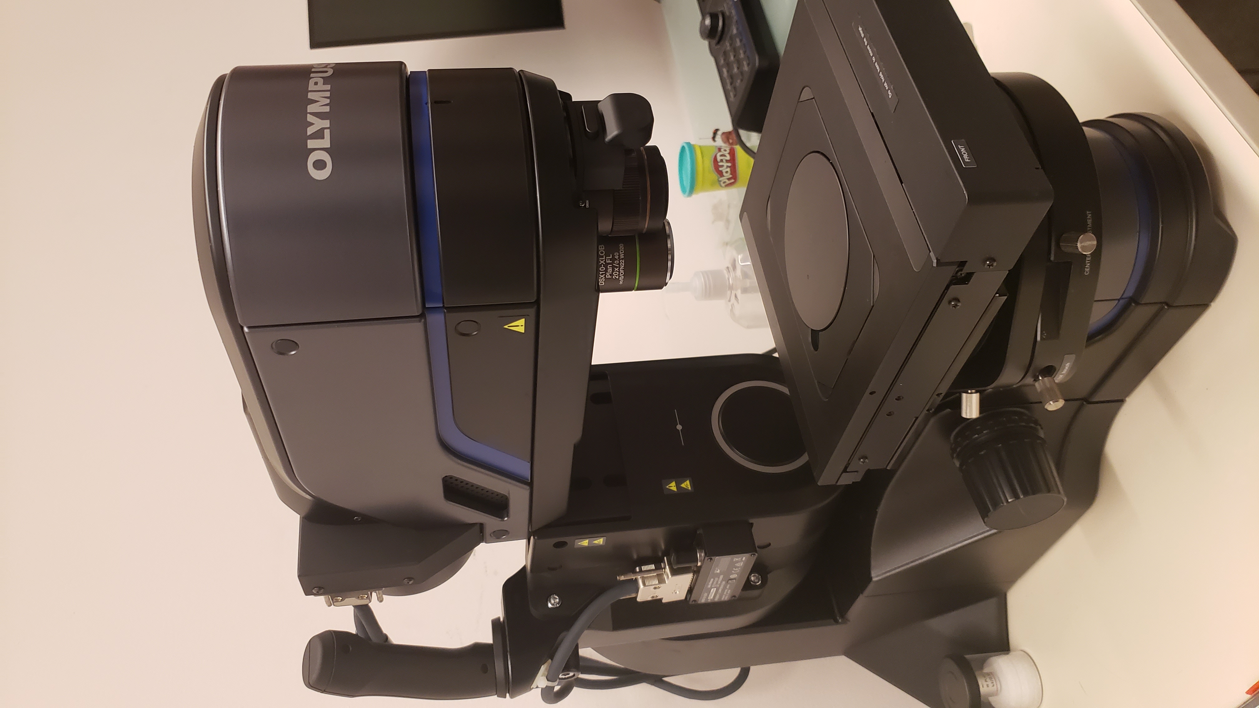
The DSX1000 microscope meets a wide range of observational and analytical needs in a single unit while improving the inspection workflow. An expanded lineup of 15 lenses covers a 20-7,000X magnification range. Users can also take advantage of the microscope’s six observation methods to observe and measure a variety of objects. For example, techniques are available to highlight irregularities on a sample surface or emphasize contours. The main unit and stage can each be freely adjusted ± 90° to accommodate samples with many shapes and to view the samples from all angles. In addition, newly developed algorithms can be used to acquire 3D images approximately ten times faster than conventional digital microscopes.
Equipment
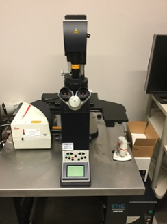
- Leica SPE Confocal
This spectral confocal features four laser lines (405 nm, 488 nm, 532 nm, and 635 nm) and tunable emission filters, with scan speeds ranging from 400 to 800 Hz. Brightfield and DIC modes are also available. We offer four ACS APO objectives on this system: 10x/0.3 dry, 20x/0.6 mixed immersion, 40x/1.15 oil, and 63x/1.3 oil. Software features include tile scan for stitching large sample areas or mark and find for choosing multiple areas of interest for imaging. Leica’s 3D viewer software allows users to save images as a volume view, a shadow view, or a maximum intensity projection. Individual snapshots or all images from a Z-stack may also be exported.
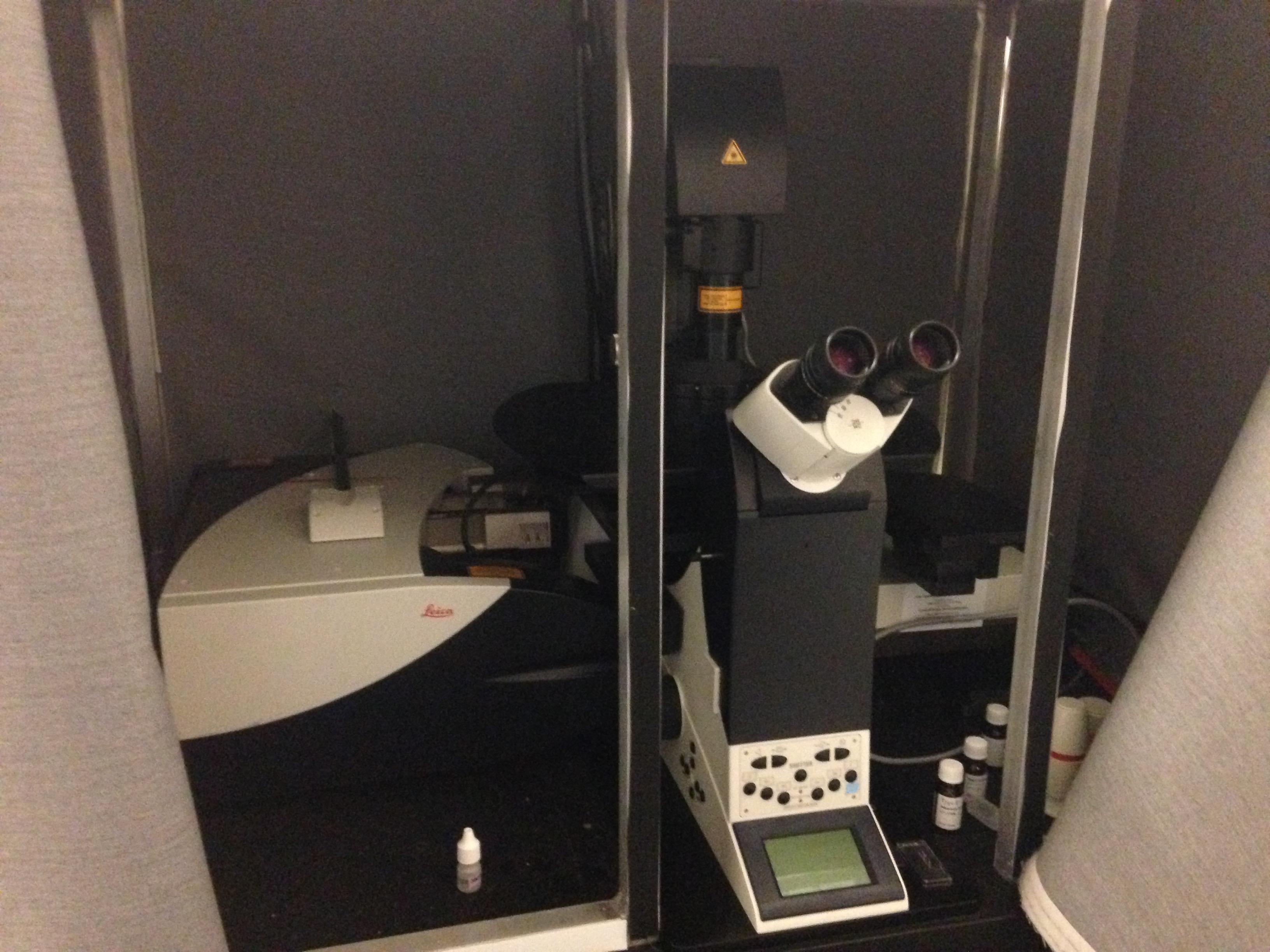
- Leica SP8X MP Confocal with Hyvolution
This high-speed multiphoton imaging system is equipped with a Ti-Sapphire laser, a tunable whitelight laser for maximum flexibility in stain choices, and high sensitivity GaAsP detectors for increased signal to noise ratio. Multiphoton imaging is less phototoxic to live cells, since infrared wavelengths are utilized. In addition, Leica’s Light Gate technology allows for flexible and adjustable time windows for collecting signals on the HyD detectors. The SP8 system also offers users access to HyVolution; this software option uses a combination of the HyD detectors and a deconvolution algorithm to achieve resolution limits of 140 nm with no changes to the sample preparation protocol.
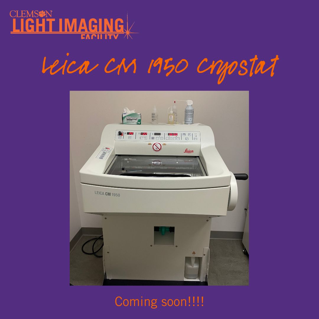
- Leica CM1950 Cryostat
The ergonomic Leica Biosystems CM1950 cryostat features Peltier cooling for rapid sample freezing and a vacuum attachment for easy clean-up. Daily defrost cycles prevent ice build-up, and built-in UV decontamination is available for anyone who may need to section potentially hazardous samples. There is a minimum 1-hour charge for use, and the hourly rate includes consumables required for cryosectioning (OCT, tissue molds, blades, freeze spray, charged slides, etc.). Researchers may choose to have a member of the CLIF staff prepare cryosections for an additional fee.
- Leica Laser Microdissection
The Leica LMD uses gravity powered specimen collection to catch user specified pieces of tissue to aid in downstream analysis work. Lasers are used in conjunction with a stylus and a touchscreen to draw the laser’s path. Most importantly, the system is contact and contamination free, and can capture homogeneous samples from heterogeneous starting samples. Users can select regions as small as a single cell, and can use living cells from cell culture. The system features objectives ranging from 1.25X to 150X, and is equipped with a Leica DFC7000T camera. Those who use the system primarily for microdissection pay a fee based on consumables, which must be purchased from CLIF. Those who wish to use the system primarily for widefield imaging must pay an hourly rate.
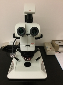
- Leica M125 Stereoscope
This ergonomic and modular routine stereoscope features a large field of view and a 10:1 zoom range. The standard configuration allows users to image with a 1.0X Plan Apo objective. A 2.0X Plan Apo lens can be installed as a custom configuration for a nominal fee. In addition to the transmitted light base, a LED ring light and LED gooseneck lights are available for optimum specimen illumination. The lighting modes include brightfield, darkfield, and Rotterman contrast. Images can be collected using a Leica DFC290HD 3 MP color camera. A third party software package, Helicon Focus, is installed on the computer and is available for generating extended depth of focus images.
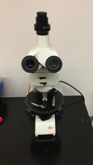
- Leica DM750P Polarized Light
This polarized light microscope allows users to investigate crystalline structures in a variety of applications. Its LED illumination lights samples homogenously and at a constant color temperature. The polarizers allow users to observe birefringence, while its strain-free optics ensure that only the sample is observed.
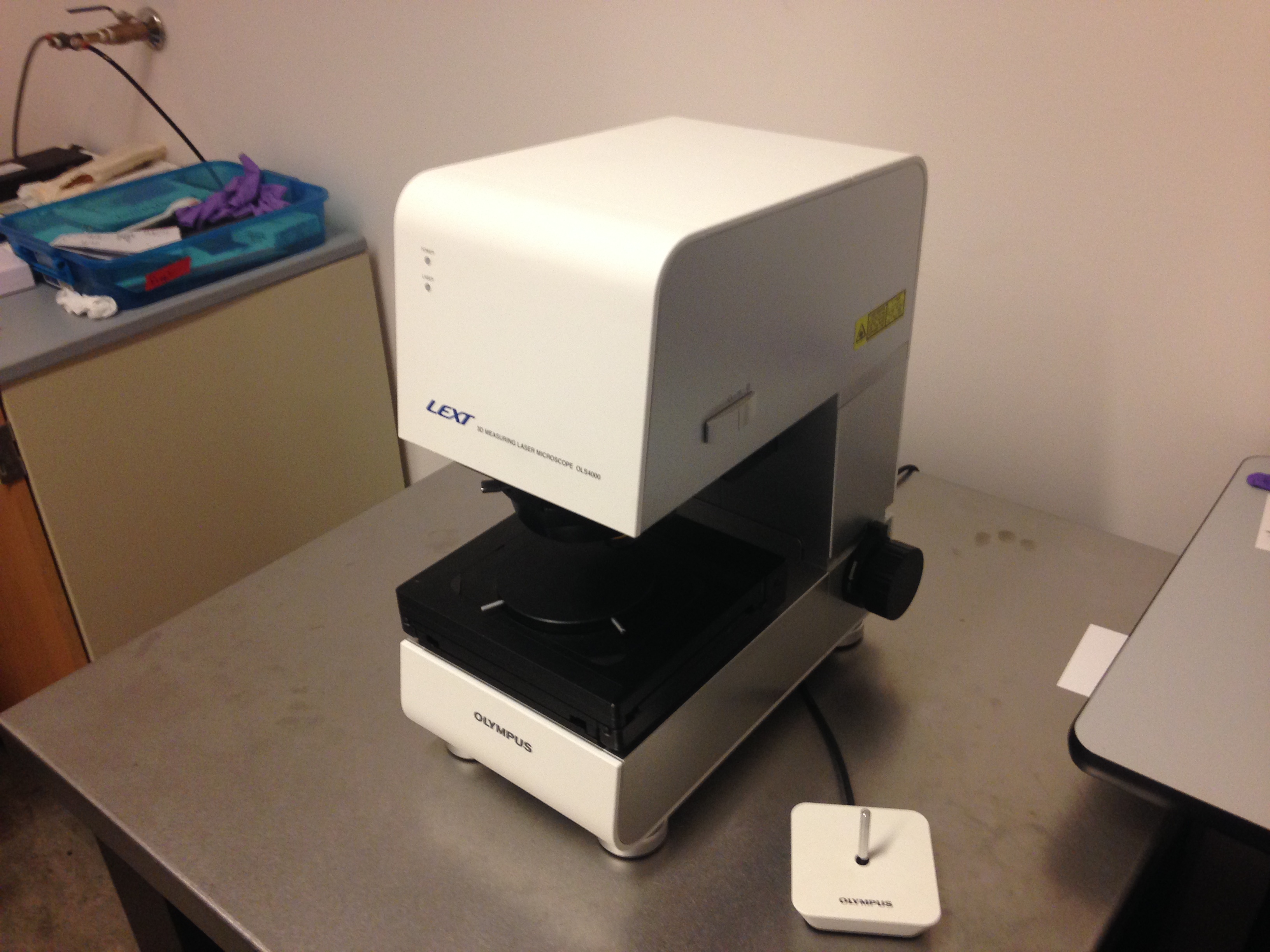
- Olympus LEXT Optical Profiler
The LEXT 3D laser measuring microscope performs non-contact roughness measurements, 3D measurements, and measurements of acute-angled specimens (slopes up to 85°). Images larger than the field of view can be connected using the stitching program. The Z-drive moves in steps less than 1nm, accuracy and repeatability guaranteed by Olympus. The system includes 5 objectives: 5x/0.15, 10x/0.30, 20x/0.60, 50x/0.95, and 100x/0.95.
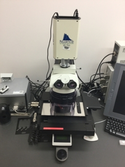
- Cytoviva Hyperspectral
The CytoViva uses patented illumination technology to collect high signal-to-noise, transmitted darkfield images of scattered light from nanoparticles. The hyperspectral imaging system captures the visible and near infrared spectrum within each pixel of the field of view. This allows for detailed spectral analysis of the scattered light from scanned materials. This “spectral library” for specific nanomaterials can be used to confirm the presence of the nanomaterials in cells, tissues, or composites.
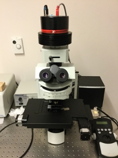
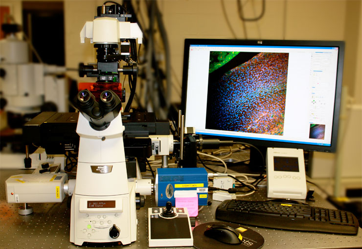
- Nikon Eclipse Ti-E
This motorized inverted microscope can be used in widefield or confocal mode. The system is equipped with a stage-top incubator with CO2, temperature and humidity control designed for imaging live cultured cells and is suitable for routine imaging and time-lapse experiments. The Perfect Focus System corrects for drift and aids stability during long term imaging. In addition to image acquisition, Nikon's NIS-Elements software can also be used for image analysis.
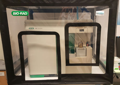
- Biorad S3E Cell Sorter
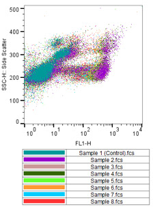
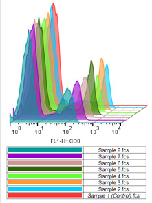
The S3e is an automated sorter with real-time technology. Features include hands-free alignment and automated drop delay calculations. The system is equipped with 2 laser lines (488 nm and 561 nm) and detectors for forward scatter, side scatter, and fluorescence. Users may perform 2-way cell sorting or may use the equipment as a flow cytometer. Generated data can be analyzed and prepared for professional presentations using our FlowJo workstation.
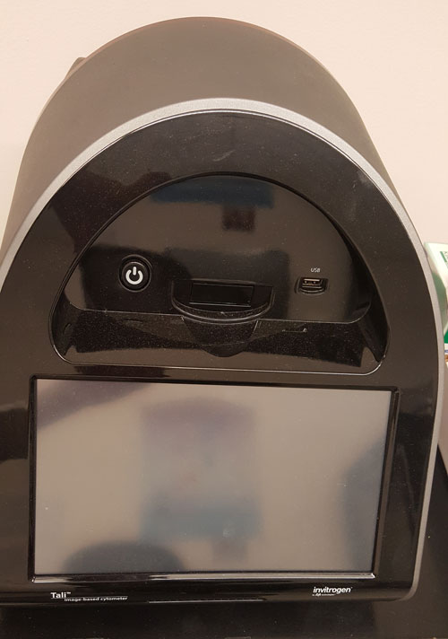
- Invitrogen Tali Image-Based Cytometer
The Tali is an imaged based cell cytometer and features three channels: brightfield, green fluorescence, and red fluorescence. The Tali has the capability to asses viability, apoptosis, oxidative stress, and cell cycle. It is compatible with a wide variety of eukaryotic cells and requires only 25 μL of sample volume, and only takes 10 seconds to 2 minutes to run a typical assay.
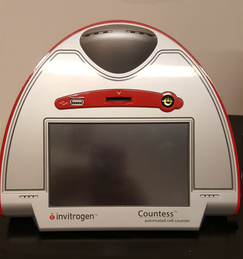
- Invitrogen Countess Cell Counter
The Countess is a cell counter that allows the option for a reusable hemocytometer. While the Countess only features one imaging channel - brightfield - live/dead counts and average cell size can be measured in approximately 30 seconds.
Software
- FlowJo Software

To further analyze Flow Cytometry Data, the CLIF has a FlowJo software license. Flowjo is an analysis software capable of visualizing .fcs data from the BioRad S3e and other cytometers. Gating procedures, cell cycle analysis, and histogram overlays are a few of the advanced features available using FlowJo. For more information and software tutorials, visit the FlowJo website.